What you do is art! Sort of.
[vc_row type=”in_container” full_screen_row_position=”middle” scene_position=”center” text_color=”dark” text_align=”left” overlay_strength=”0.3″ shape_divider_position=”bottom” bg_image_animation=”none”][vc_column column_padding=”no-extra-padding” column_padding_position=”all” background_color_opacity=”1″ background_hover_color_opacity=”1″ column_link_target=”_self” column_shadow=”none” column_border_radius=”none” width=”1/1″ tablet_width_inherit=”default” tablet_text_alignment=”default” phone_text_alignment=”default” column_border_width=”none” column_border_style=”solid” bg_image_animation=”none”][vc_column_text]A career in a pathology lab may not be for the squeamish, it’s true. Not everyone appreciated junior high science class when it came to dissecting worms and frogs. But zoom in a little further, and what’s on that slide might be quite beautiful – even to those with the most sensitive stomachs.
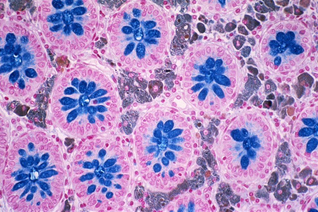
Among the inventory of duvet covers and nursery décor on FineArtAmerica.com is a section of “microscapes” from the large intestine and other areas of the human body. This beautiful pink and blue photo depicts Melanosis coli, which I learned is benign pigmentation disorder in the wall of the colon (look for the brown spots). I never thought I may want a photo of someone’s colon disorder above my mantel, but for $45, I could have quite the conversation piece in my living room.
 I highly recommend browsing all the microscapes on that website, and at the larger collection on Science Photo Library’s website. There are all kinds of body systems represented. You may run across a few less attractive images, but if you work in a lab, you’ve probably seen worse! And either way, it helps give a new perspective on what you do, and how far science has come.
That isn’t the only means of connecting pathology to art. This article from CAP TODAY describes a The Healing Art of Pathology, a book they published containing art by pathologists and other healthcare professionals, as well as patients and family members. It is available on CAP’s website for a discount for members, or on Amazon for those Prime members out there.
To see that there is a market for these images may help you gain a renewed appreciation for what you do and how far science has come. If nothing else, at least you can get a little birthday gift inspiration for the lab professional who has everything.
Photo: Melanosis coli, light micrograph by CNRI/Science Photo Library. Included in this blog with permission from Science Photo Library[/vc_column_text][divider line_type=”Full Width Line” line_thickness=”1″ divider_color=”default”][vc_column_text] Michelle Austin
Michelle Austin joined the Psychē Systems support team in 2018. Michelle has worked in the medical field for over seven years. Previously, she held the position of an Application Specialist in a clinical lab setting. She has a bachelor’s degree in English with a minor in Anthropology. She is currently in the throes of parenting a toddler and severely lacking in free time. In those fleeting spare moments, Michelle is trying to learn how to speak Spanish.[/vc_column_text][/vc_column][/vc_row]
