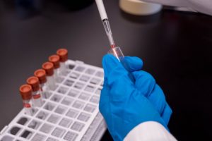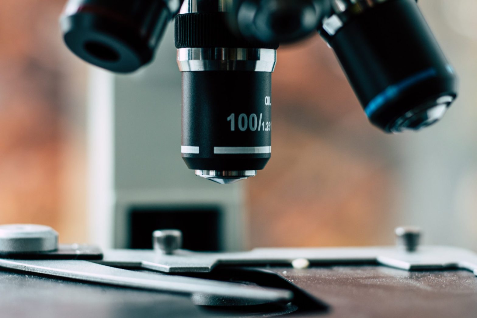

Cytogenetics is the microscopic study and analysis of chromosomes within the nucleus. It can be used when testing samples for genetic disease. The analysis involves studying the structure and number of chromosomes in order to identify changes. Some of the changes in chromosomes that may influence genetic disease involve missing, broken, or extra chromosomes. The types of samples studied are usually samples that come from blood, tissue, or bone marrow.
Cytogenetics is a useful tool in identifying and treating genetic diseases early on. Here are some of the techniques and methodology between cytogenetics and genetic diseases.
Distinguishing Individual Chromosomes
In order to distinguish individual chromosomes, a technique called G-banding is used. This technique was developed in 1971 by researchers Margery Shaw and Maximo Drets. In order to differentiate unique human chromosomes, the researchers discovered using Giemsa dye to identify the banding patterns, now referred to as G-bands. In order to reach the Giesma dye phase, the researchers had to take human lymphocytes and fibroblasts that were going through mitosis and then expose them to colchicine. After this exposure, they were permeabilized by being exposed to hypotonic conditions. Once permeabilization is complete, they are exposed to methanol and acetic acid. After this exposure, they were flame dried, treated with sodium hydroxide, and then finally the Giesma dye was introduced in order to identify the G-bands.
The benefits of the G-banding technique allow researchers to identify chromosome numbers and structures. This lets researchers observe restructuring or deletion of chromosomes, thus benefiting the research and identification of genome-related diseases. The G-band technique also provides scientists the ability to identify individual chromosomes that have been deleted or translocated. The identification of individual types of chromosomes and translocations or deletions allows the opportunity to diagnose specific diseases with precision.
Routine Karyotyping to Identify Genetic Disease
This is a form of chromosomal identification and analysis. It also commonly involves the usage of dyes, most frequently the previously mentioned Giemsa dye. A routine karyotyping involves organizing the band into a karyogram of 23 pairs. Scientists can identify abnormalities such as translocations, deletions, or an incorrect number of chromosomes. However, routine karyotyping is limited to identifying only approximately three megabases. Routine karyotyping can identify genetic diseases such as Turner Syndrome and Down Syndrome.
Comparative Genomic Hybridization
Comparative Genomic Hybridization, or CGH, is a useful cytogenetic diagnostic tool when a diagnosis is unknown. This method grants the ability to efficiently scan an entire genome. During the scan, chromosomal abnormalities can be detected.
This method works by detecting chromosomal copy number variants. Scientists can do this with CGH without the need for cell culturing. CGH also uses two chromosomes, a test sample, and a control sample. They are both dyed, denatured, and mixed together. The differences in the dye intensity are compared and contrasted between the test chromosome and control chromosome to detect abnormalities. The resolution of CGH is approximately up to 10 megabases.
Similar to CGH is another method known as Array Comparative Genomic Hybridization, or aCGH. This method uses a similar technique; however, the resolution is higher than CGH. This happens with the usage of microarrays. Small sections of chromosomes are identified for scanning and are then immobilized without altering the proteins. The drawback of this method is that it cannot detect balanced inversions or translocations.
Fluorescent in situ Hybridization
Fluorescent in situ hybridization, or FISH, is a technique used to identify both acquired and congenital diseases. One of the benefits of FISH is that it has a significantly higher resolution than routine karyotyping. FISH is a commonly used diagnostic tool in cytogenetics.
FISH locates the presence or absence of different types of chromosomes and strands of DNA by using fluorescent probes. Both the chromosome and probe must first be denatured in order to break the hydrogen bonds in the DNA. Denaturing allows hybridization to occur after the two samples mix with each other. After they are mixed, researchers can use a microscope to identify new hydrogen bonds created with the fluorescent probes.
FISH is most useful in many pediatric situations to detect chromosomal abnormalities. Different types of cancers and diseases that are prevalent in children can be detected using this method.
Multiplex Ligation-dependent Probe Amplification Assay
Multiplex Ligation-dependent Probe Amplification, or MLPA, is a technique used to identify a group of genetic diseases on certain chromosomes. This technique detects deletions or duplications of certain DNA, as well as the detection of abnormal DNA methylation. MLPA is useful in detecting prenatal abnormalities, as well as different types of cancer.
This process involves denaturing two strands of DNA and then adding two MLPA probes to induce hybridization. After hybridization, ligation and amplification occur. This allows the DNA copies to be analyzed and measured. Amplification allows the DNA strands to be separated based on their length. This is performed through capillary electrophoresis. The data obtained is then compared to reference samples to calculate the probe ratio. This will reveal the copy number of a gene.
Reviewing Chromosomes
When chromosomes are prepared in a karyotype, it is referred to as a karyogram. In these karyograms, chromosomes are suspended in a horizontal axis. The specific placement of chromosomes in a karyogram allows researchers to diagnose the size, or morphology, of the chromosome. The majority of karyograms have lower resolutions; however, they are still useful in detecting a number of different abnormalities. The completion of the Human Genome Project greatly aids cytogenetic research.
Contact Psychē Medical Laboratory Software
In the world of cancer and genetic disease treatment, the tools used for cytogenetic research continue to advance. In order to meet the medical needs of patients, diagnostic labs must be sure to include updated and advanced cytogenetic techniques and methodology in their portfolio.
Contact Psychē Medical Laboratory Software to discuss your options. We are happy to help you improve the efficiency of your diagnostic lab.